A 58-year-old man is to have an ankle arthrodesis for post-traumatic arthritis. The hindfoot and midfoot joints are mobile and free of arthritis.
In which position should the talus be fused to the tibia?
How sure are you?
Trauma · Free article
Ankle fractures for FRCS (Tr & Orth): Weber and Lauge-Hansen, judging stability, BOAST 12, the AIM trial, the syndesmosis, posterior malleolus and diabetes.
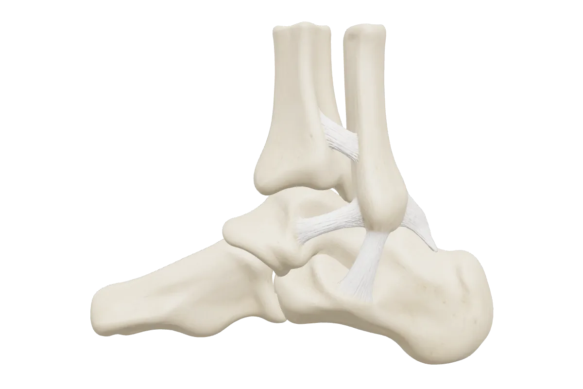
The talus sits in a mortise formed by the tibial plafond and the medial and lateral malleoli. It is held there by three groups of ligaments: the deltoid ligament medially, whose deep part is the main restraint to lateral talar shift; the lateral ligaments (anterior talofibular, calcaneofibular and posterior talofibular); and the syndesmosis, made of the anterior and posterior inferior tibiofibular ligaments, the interosseous ligament and the inferior transverse ligament.
Think of a ring of bone and ligament around the talus. A single break leaves the ring stable; two breaks, for example a fibular fracture with a deltoid rupture, allow the talus to shift. Ramsey and Hamilton (1976) showed that a millimetre of lateral talar shift sharply reduces the tibiotalar contact area, which is why talar shift is not accepted.
| Weber | Fibular fracture | Syndesmosis | Usual mechanism |
|---|---|---|---|
| A | Below the syndesmosis, often transverse | Intact | Supination–adduction |
| B | At the level of the syndesmosis, usually spiral | May be partly injured | Supination–external rotation |
| C | Above the syndesmosis | Disrupted | Pronation–external rotation or pronation–abduction |
Lauge-Hansen (1950) is a mechanistic system: the first word is the position of the foot at injury and the second the direction of the deforming force. Each type has stages that occur in sequence, so the stage predicts which structures are injured.
| Type | Sequence of injury |
|---|---|
| Supination–external rotation (commonest) | I anterior inferior tibiofibular ligament → II spiral fibular fracture at the syndesmosis (anteroinferior to posterosuperior) → III posterior inferior tibiofibular ligament or posterior malleolus → IV medial malleolus fracture or deltoid rupture |
| Supination–adduction | I transverse avulsion of the lateral malleolus below the joint → II vertical medial malleolus fracture, sometimes with impaction of the medial plafond |
| Pronation–external rotation | I medial malleolus fracture or deltoid rupture → II anterior inferior tibiofibular ligament → III high spiral fibular fracture above the syndesmosis → IV posterior inferior tibiofibular ligament or posterior malleolus |
| Pronation–abduction | I medial malleolus fracture or deltoid rupture → II syndesmotic ligaments → III transverse or comminuted fibular fracture above the syndesmosis |
BOAST 12 asks for diabetes, peripheral neuropathy, peripheral vascular disease and other comorbidities to be documented at presentation because they change the outcome. Patients with diabetic neuropathy have more wound breakdown, infection, loss of fixation and Charcot neuroarthropathy. Many surgeons use stronger constructs (for example extra syndesmotic fixation or a hindfoot nail in frail patients), protect weight bearing for longer and follow them up more closely.
Test yourself
A 58-year-old man is to have an ankle arthrodesis for post-traumatic arthritis. The hindfoot and midfoot joints are mobile and free of arthritis.
In which position should the talus be fused to the tibia?
Injury on both sides of the ring around the talus, allowing it to shift. Typically a fibular fracture with a medial malleolar fracture or a deep deltoid ligament rupture, seen as a widened medial clear space.
No. An isolated Weber B fracture with an intact deltoid ligament, confirmed by a normal medial clear space on weight-bearing or stress views, is stable and is treated in a boot or cast with weight bearing as tolerated.
A proximal fibular fracture with disruption of the ankle syndesmosis and the interosseous membrane, and a medial ankle injury. It is a pronation–external rotation injury and is treated by stabilising the syndesmosis.
Yes, if the reduction can be held. BOAST 12 accepts a close contact cast for patients over 60, and the AIM trial found function equivalent to surgery at six months, with fewer wound complications but more malunion.
When it is unstable on intraoperative testing after the malleoli have been fixed, using a hook test or an external rotation stress test under fluoroscopy. It is held with screws or a suture-button device after careful reduction.
Written for revision in the FRCS (Tr & Orth) style under our editorial standards. It is not clinical advice: always confirm against current guidance. Spotted an error? Tell us.
Trauma
Hip fractures for FRCS (Tr & Orth): Garden and Pauwels, femoral head blood supply, NICE CG124 and its 2023 THR criteria, cemented implants, screw or nail.
ReadTrauma
The BOAST open fracture standard for FRCS (Tr & Orth): antibiotics within an hour, debridement at 12 or 24 hours, cover within 72 hours, and Gustilo–Anderson grading.
ReadBasic science
Primary and secondary bone healing, the stages of callus, Perren’s interfragmentary strain theory, and how to choose absolute or relative stability for a fracture.
ReadDrill trauma with curriculum-mapped questions, rate your confidence, and watch the gap close on your dashboard.