Paediatrics · Free article
Physeal fractures: Salter–Harris, growth arrest and the transitional fractures of the ankle
Physeal fractures for FRCS (Tr & Orth): zones of the physis, Salter–Harris I–V and Rang VI, growth arrest, bar resection, Tillaux and triplane fractures.
The physis
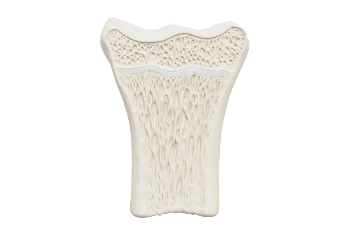
The physis is a disc of cartilage between the epiphysis and the metaphysis. Its zones are arranged from the epiphyseal side towards the metaphysis, and each has a different job and a different mechanical strength.
- Resting (reserve) zone
- Scattered chondrocytes in abundant matrix, next to the epiphysis. Stores nutrients and supplies cells to the zone below.
- Proliferative zone
- Chondrocytes divide and stack into columns. This is where longitudinal growth is generated; achondroplasia is a defect of this zone.
- Hypertrophic zone
- Cells enlarge, then die as the matrix around them calcifies (the zone of provisional calcification). It has the least matrix and is mechanically the weakest. Rickets is a failure of provisional calcification, and slipped capital femoral epiphysis occurs through this zone.
- Metaphysis
- Vascular invasion lays down primary spongiosa on the calcified cartilage. Osteomyelitis and scurvy are metaphyseal diseases.
Around the edge, the groove of Ranvier supplies cells for growth in width and the perichondral ring of LaCroix gives mechanical support. The germinal layer is nourished from the epiphyseal side, so where the epiphysis is entirely intra-articular (the proximal femur and the proximal radius), separation can cut off its blood supply.
The Salter–Harris classification
| Type | Fracture line | Notes |
|---|---|---|
| I | Straight through the physis | Radiographs may be normal; suspect it when the physis is tender. Slipped capital femoral epiphysis is an atraumatic type I injury |
| II | Through the physis and out through the metaphysis | The commonest type. The metaphyseal (Thurston Holland) fragment lies on the compression side, where the periosteum is intact and helps reduction |
| III | Through the physis and out through the epiphysis | Intra-articular; crosses the germinal layer |
| IV | Across the metaphysis, physis and epiphysis | Intra-articular; displacement can let bone bridge the physis and form a bar |
| V | Crush of the physis | Often recognised only in retrospect, when growth arrest appears |
| VI (Rang) | Injury to the perichondral ring at the edge of the physis | Often from a burn, an abrasion or a lawnmower; a peripheral bar causes angular deformity |
What predicts growth disturbance
- Type. Types III, IV and V are more likely to cause arrest than I and II, but the type is not the whole story.
- Which physis. The undulating distal femoral and proximal tibial physes interlock with the metaphysis, so a fracture shears across the ridges and damages germinal cells; arrest is common after distal femoral physeal fractures, even type II injuries. These physes also contribute most of the growth of the lower limb, so arrest costs more length: growth happens near the knee and away from the elbow.
- Age. A younger child has more growth to lose, but also more capacity to remodel.
- Energy and displacement. High-energy, comminuted and widely displaced fractures fare worse.
- Treatment. Repeated or late forceful manipulation and threaded implants across the physis can cause arrest themselves.
Remodelling is greatest close to the physis, in the plane of movement of the nearest joint, and in younger children. Rotational malunion does not remodel.
Managing physeal fractures
- Types I and II. Gentle closed reduction early, then a cast. In young children some residual angulation in the plane of joint movement can be accepted because it will remodel. If the fracture is unstable, smooth Kirschner wires may cross the physis.
- Types III and IV. Anatomical reduction of the joint surface and the physis, often open. Screws are placed parallel to the physis, within the epiphysis or the metaphysis, without crossing it.
- Follow-up. Fractures at high risk of arrest are followed with radiographs until normal growth is confirmed. A Park–Harris growth arrest line that runs parallel to the physis is reassuring; one that converges towards the physis points to a bar.
Physeal bars
A bony bridge across the physis tethers growth. A central bar causes shortening and a tented physis; a peripheral bar causes angular deformity. MRI or CT maps its size and position. Langenskiöld’s criteria for bar resection with interposition of fat or cement are a bar involving less than half of the physis in a child with at least two years of growth remaining. Otherwise the arrest is completed by epiphysiodesis (with contralateral epiphysiodesis if the length discrepancy would be significant), and deformity or shortening is corrected by osteotomy or lengthening.
Transitional fractures of the distal tibia
In adolescence the distal tibial physis closes over about 18 months, starting centrally, then medially and finally laterally. While the anterolateral part is still open, external rotation injuries produce two characteristic fractures.
- Tillaux fracture. A Salter–Harris III fracture of the anterolateral epiphysis, avulsed by the anterior inferior tibiofibular ligament.
- Triplane fracture. A Salter–Harris IV pattern in three planes: sagittal through the epiphysis, axial through the physis and coronal through the metaphysis. It may have two, three or four parts.
- Imaging and treatment. CT defines the fragments. Articular displacement of more than about 2 mm after closed reduction is an indication for open or percutaneous reduction and fixation.
- Little remaining growth. Because the physis is already closing, growth arrest is less of a concern than joint congruity.
In younger children, a Salter–Harris III or IV fracture of the medial malleolus is different: a bar can form across the open medial physis and cause progressive varus deformity, so these need anatomical reduction and careful follow-up.
Sites that need respect
- Distal femur
- High risk of growth arrest after any type. Hyperextension injuries that displace the epiphysis anteriorly endanger the popliteal vessels.
- Proximal tibia
- The popliteal artery is tethered near the trifurcation and can be injured by posterior displacement of the metaphysis; watch for compartment syndrome.
- Proximal humerus
- Contributes most of the growth of the humerus and remodels remarkably, so considerable displacement can be accepted in younger children.
- Distal radius
- The commonest site of physeal fracture and very forgiving. Avoid late remanipulation.
Frequently asked questions
Which zone of the physis do most fractures pass through?
The hypertrophic zone, near the zone of provisional calcification, where there is least matrix. The resting and proliferative layers stay with the epiphysis, which is why types I and II usually grow normally.
Which Salter–Harris type is most common?
Type II, with a fracture through the physis that exits through the metaphysis, leaving a Thurston Holland fragment on the compression side.
When can a physeal bar be resected?
When it involves less than half of the physis and the child has at least two years of growth remaining (Langenskiöld). The bar is excised and the gap filled with fat or cement to stop it reforming.
What is a Tillaux fracture?
A Salter–Harris III fracture of the anterolateral distal tibial epiphysis in an adolescent, avulsed by the anterior inferior tibiofibular ligament while the lateral part of the physis is still open.
Why do distal femoral physeal fractures cause growth arrest so often?
The physis is undulating, so a fracture shears across its ridges and damages germinal cells, and it contributes much of the growth of the leg. Arrest is common even after type II injuries.
References
- Salter RB, Harris WR. Injuries involving the epiphyseal plate. J Bone Joint Surg Am 1963;45(3):587–622.
- Rang M. The Growth Plate and Its Disorders. Edinburgh: Livingstone; 1969.
- Peterson HA. Physeal fractures: Part 3. Classification. J Pediatr Orthop 1994;14(4):439–448.
- Langenskiöld A. Surgical treatment of partial closure of the growth plate. J Pediatr Orthop 1981;1(1):3–11.
- Basener CJ, Mehlman CT, DiPasquale TG. Growth disturbance after distal femoral growth plate fractures in children: a meta-analysis. J Orthop Trauma 2009;23(9):663–667.
Further reading
- Radiopaedia — Salter–Harris classification — Radiographic examples of each type
Written for revision in the FRCS (Tr & Orth) style under our editorial standards. It is not clinical advice: always confirm against current guidance. Spotted an error? Tell us.
Related articles
Basic science
Fracture healing and Perren’s strain theory: absolute and relative stability
Primary and secondary bone healing, the stages of callus, Perren’s interfragmentary strain theory, and how to choose absolute or relative stability for a fracture.
ReadTrauma
Ankle fractures: Weber, Lauge-Hansen and the question of stability
Ankle fractures for FRCS (Tr & Orth): Weber and Lauge-Hansen, judging stability, BOAST 12, the AIM trial, the syndesmosis, posterior malleolus and diabetes.
ReadTrauma
Open fractures: the BOAST standard, timings and the Gustilo–Anderson classification
The BOAST open fracture standard for FRCS (Tr & Orth): antibiotics within an hour, debridement at 12 or 24 hours, cover within 72 hours, and Gustilo–Anderson grading.
ReadTurn reading into marks
Drill paediatrics with curriculum-mapped questions, rate your confidence, and watch the gap close on your dashboard.